Prenatal diagnosis of congenital heart defects using unique magnetic resonance imaging method
Most often a congenital heart defect is suspected during a routine fetal ultrasound examination. But sometimes suspicion arises late in pregnancy, when an ultrasound examination may be more challenging. In these cases, it can be difficult to be confident about the diagnosis.
Not taking any risks, medical staff often prepare for a delivery where the baby is born with a heart defect, even though it might not always be necessary.
“This could mean that the parents-to-be need to stay at a highly specialised hospital before delivery, and also give birth at that hospital instead of at their home hospital”, says Erik Hedström, Consultant at Skåne University Hospital and Associate professor in the Lund Cardiac MR Group, Lund University and Skåne University Hospital.
Diagnostic images by MRI
The Lund Cardiac MR Group has developed a unique MRI method that provides high-quality images and movies of the beating fetal heart. The images and movies of the fetal heart can, amongst other things, be used to assess cardiac function. This helps in cases when a routine ultrasound does not provide enough information.
“When we first introduced the possibility of imaging the fetal heart, it took us one to two weeks after the examination to actually reconstruct images to view. Now we have managed to get the images directly on screen during the examination. The advantage is that we can tell directly and with certainty whether the fetus has a congenital heart defect or not, and how serious it is in that case. This means that the parents get information they can trust, and clinicians can prepare for the delivery in the best possible way”, says Erik Hedström.
Their son's heart defect was clarified

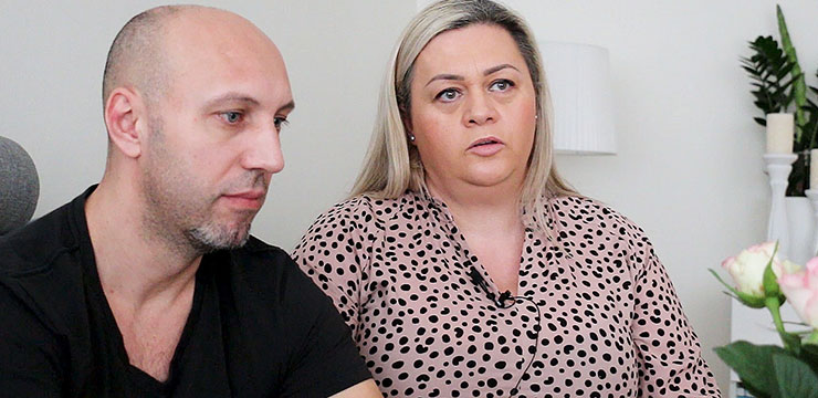
Fatos Voca and Amelika Voca.
Amelika Voca was one of the first patients in Sweden to be examined with the MRI method. The suspicion that her son Lian had a congenital heart defect was raised when she was seven months pregnant.
“I had a routine ultrasound examination. Suddenly the physician became very serious and said that he had found something. He and his colleagues looked at it together and said that they suspected a heart defect, but they didn’t know for sure. Living in uncertainty was incredibly difficult”, she says.
To get more detailed information about Lian's heart, Amelika Voca was examined with the unique MRI method at Skåne University Hospital. It then became clear that Lian would in fact be born with a serious congenital heart defect.
“Those were devastating news, but in a way Lian’s father and I felt relieved that at least we knew for sure what to expect. After Lian was born, there was only one alternative: surgery. Otherwise, he wouldn’t have made it. Today he is a happy and mischievous three-year-old”, Amelika Voca says.
Further improvement
Further development of the MRI method is ongoing. The goal is to improve image resolution to be able to examine smaller structures, and to speed up image acquisition to simplify the examination for the pregnant woman.
“Routine ultrasound and this MRI method complement each other. There are however cases when we do not reach a conclusive diagnosis with any of the methods. We continue the development to also in those cases clarify the information to the parents-to-be so that they can prepare already before delivery what to expect, increase patient safety, and improve utilisation of healthcare resources.
Facts: This is how the method works
- Simplified, an image from an MRI scanner consists of many lines collected one at a time. Since the heart is constantly beating, it is extremely difficult to acquire images of the beating heart, even more so for the fetal heart, which beats quickly at about 140 beats per minute. Furthermore, the fetal heart is only about five centimetres in diameter at term.
- In adults, an ECG is used to sort the collected lines at the right time during the heartbeat, and thus get a movie of the beating heart. As the fetal ECG signal is very weak compared with the mother’s, this is not possible in fetal heart examinations.
- To overcome this, the Lund Cardiac MR Group utilises an MR-compatible Doppler Ultrasound device developed at Hamburg University by Fabian Kording, PhD, and coworkers. This device detects the fetal heart beats and provides a signal for the MR scanner to sort the collected lines and make a movie of the beating fetal heart.
- By further refining advanced MRI methods, Skåne University Hospital can depict fetal hearts and their function using a standard MRI scanner.
Source: Erik Hedström, Consultant at Skåne University Hospital and Associate Professor in the Lund Cardiac MR Group, Lund University and Skåne University Hospital.

