Unique method provides faster postoperative recovery for children with congenital heart defects
Congenital heart defects are the most common congenital malformations and are the leading cause of mortality from birth defects. For those requiring complex surgeries and transcatheter interventions, a predictive computer simulation of treatment outcome would be invaluable.
Currently, simulations using computational fluid dynamics (CFD) offer the possibility to analyse the outcome. However, clinical use of CFD is still limited. A major reason is the method’s complexity, which includes need for advanced software, time-demanding analysis, and multiple interaction between clinicians, medical physicists, and engineers.
Thanks to a novel, leaner method of CFD, it now takes five minutes instead of up to 40 hours to measure how pulmonary flow is affected during certain surgical interventions. The method has been developed by Petter Frieberg at the Lund Cardiac MR Group at Lund University, in close collaboration with Petru Liuba at the Paediatric Cardiac Centre at Skåne University Hospital. Several patients with a so-called univentricular heart malformation have already benefited greatly from the method.
“Thanks to the new method, we are now able to guide complex cardiac interventions to obtain the best postoperative outcome. Each patient with congenital heart defect is unique in terms of cardiac anatomy and physiology. In transcatheter interventions, treatment decision making is sometimes based on data obtained in the beginning of the intervention. Using this method, we can also decide during the same procedure which treatment option we should choose. This is important since all paediatric cardiac interventions typically require general anaesthesia, avoiding thus unnecessary reinterventions, complications due to reinterventions and excessive resource utilization. This means great benefits for the child, the family and for healthcare in general”, says Petru Liuba, responsible for cardiac catheterization at the Paediatric Heart Centre at Skåne University Hospital and senior lecturer at Lund University.

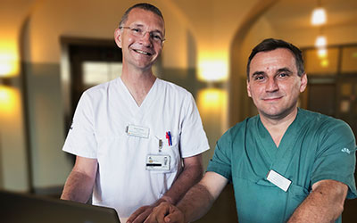
Petter Frieberg and Petru Liuba.
A less invasive intervention
The method is already used in both planning and guiding some of the cardiac operations at Skåne University Hospital. The new method is used, among other things, in procedures on children born with single ventricle.
“Heart defects in children vary from patient to patient, even among children with the same type of congenital heart defect. Therefore, each cardiac intervention requires both individualized assessment and careful discussion with other colleagues with various expertise, including cardiac imagers, anaesthesiologists and radiologists. This is why and how the joint work with my colleagues Petter Frieberg and Marcus Carlsson from the Lund Cardiac MR group started. Dr Frieberg's contribution to developing this method has been of paramount importance”, Petru Liuba says.
“With the new method, it only takes a few minutes for us to evaluate the consequences different interventions will have on the child, even during an ongoing cardiac intervention”, he continues.
After complex interventions, patients may need to stay in the hospital up to several months. Especially in patients with univentricular heart requiring reinterventions, it can take months or even years before clinical improvement occurs.
“We have had cases where, thanks to the new technology, we have been able to make more gentle, less invasive interventions. Children were thus able to leave the hospital a few days after the intervention, which has an enormous significance for the child's quality of life and the family's situation in general”, Petru Liuba continues.
Innovative work
Skåne University Hospital has shortened the calculation time by combining the traditional flow analysis software with an advanced simulation program used by engineers in design departments. Based on a 3D model of the blood vessels, a computer calculation of the pulmonary flow is then performed.
“We are the first hospital in the world to integrate these two programs and to place them in a clinical environment. We hope to soon be able to offer a reliable support system that can be used clinically, both before and during ongoing surgeries, says Petter Frieberg, physician and engineer at the Clinical Physiology and Nuclear Medicine department at Skåne University Hospital, and doctoral student at the Lund Cardiac MR Group at Lund University.
Retrospective study
The researchers have conducted a retrospective study that shows that the method is safe in relation to the established programs that are otherwise used. The study is based on 15 already performed surgeries on children who have undergone single ventricle heart surgery. By comparing results from both the new and the more traditional method with patient measurements obtained with an MRI camera, the researchers have concluded that the results are very similar.
The researchers' conclusions are that the new technology is just as good at predicting how pulmonary flows are affected as the more traditional technology used today, but considerably faster.

