Thousands of patients avoid chest X-rays thanks to choosing wisely

The emergency care and internal medicine division is pioneering the adoption of "Choosing Wisely" in Swedish healthcare. For nearly a decade, staff have carefully scrutinised treatments that offer limited value to patients. Initially modelled after the American concept, they now develop their own approaches—such as reducing chest X-rays.
"It all started with frustration among staff and questioning why we ordered so many X-rays, especially when we knew that their clinical value was often limited," says Bitte Zetterman, development officer and nurse.
Bitte Zetterman and consultant Hannes Hartman describe how a suspected case of pneumonia often triggers a "package" of diagnostic steps. This can quickly lead to standard daily tests, hospitalisation for observation, antibiotics, and, frequently, a chest X-ray—one of the most common imaging studies.
"We book the X-ray, and the patient often waits for it, sometimes overnight or until the following day. This requires transportation and additional waiting for results before we can proceed. The results are rarely conclusive for diagnosing pneumonia and may have already become irrelevant in the overall treatment process. So naturally, we started asking if this approach was necessary," says Hannes Hartman.
Patient-centred smart choices
The solution has been to move away from standard protocols in favour of a more patient-centred approach. Rather than rigidly following a care program designed for the "average" patient, healthcare professionals are encouraged to think critically: "What adds value for this particular patient?"
"A lot of this involves doing the groundwork—getting away from the screen and actually examining the patient in front of you. You need to look, feel, and listen to make an informed decision alongside the patient," Hannes Hartman explains.
14,000 fewer tests per month
This approach has previously led to a 43% reduction in blood transfusions and a decrease of approximately 14,000 lab tests ordered per month. The reduction in chest X-rays was further enabled by a large-scale implementation of point-of-care ultrasound (POCUS).
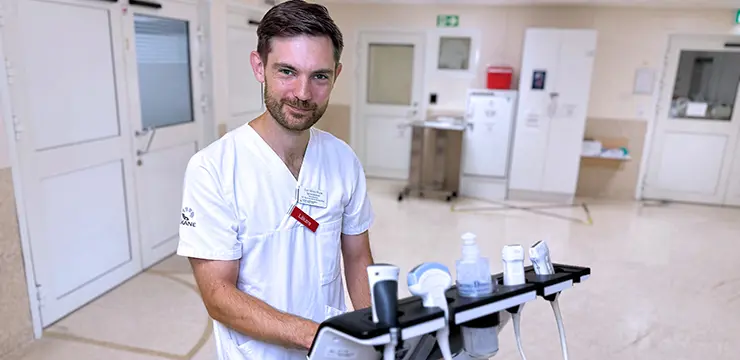
"POCUS provides us with additional clinical data to support our assessments. For example, a physician can detect fluid in the pleural cavity, potentially avoiding the need for an X-ray. Similarly, a nurse can use ultrasound guidance to place an intravenous line without requiring the patient to be transported elsewhere," Hannes Hartman adds.
Research supports new methods
The decision to replace certain X-rays with point-of-care ultrasound was preceded by several internal evaluations of scientific evidence. Researchers were tasked with comparing the methods and concluded that for specific clinical questions, ultrasound was as effective as X-ray.
"At the turn of the year, we decided to prioritize lung ultrasound and completely discontinue the practice of ordering routine supine chest X-rays, which rarely provide useful information. Our goal was to halve the number of chest X-rays over the course of the year. By April, we had already reached this target, and the numbers have continued to decline since," says Hannes Hartman.
Bitte Zetterman notes that the introduction of point-of-care ultrasound has not only made it easier to forgo X-rays but has also significantly shortened the time from patient admission to diagnosis. "The wait time and transportation disappear. Patients receive a pain-free examination on-site, avoiding radiation exposure. The results are immediate, allowing us to decide on the next step in the treatment process without delay," she says.
"And healthcare staff get more time with the patient," Hannes Hartman adds. "No patient feels as thoroughly examined as when they’ve had a ten-minute ultrasound examination by their doctor. Additionally, radiology departments can focus on more urgent cases, and in the long run, our resources go further."


