

Brain tumour patients successfully treated with laser interstitial thermal therapy inside an MRI scanner
All patients were diagnosed with glioblastoma multiforme (GBM), the most common and most aggressive form of brain tumour. The purpose of the study was to investigate whether MRI-guided LITT for patients with brain tumours is safe and feasible.
The results of the study were presented on 11–15 October at the Congress of Neurological Surgeons Annual Meeting in Los Angeles, USA.
Treating tumours in difficult-to-reach areas
“This study shows that we can now successfully treat patients with brain tumours located in areas that were previously considered inoperable. In the past, surgery carried too high a risk using traditional open techniques,” says Peter Siesjö, senior consultant in neurosurgery at Skåne University Hospital, adjunct professor at Lund University, and principal investigator of the study.
The procedure involves inserting a thin laser catheter into the brain and targeting the tumour, which is then destroyed by heat. The remnants of the tumour are naturally cleared away by the body’s waste disposal system. The operation is performed while the patient is inside an MRI scanner, allowing surgeons to monitor the treatment in real time through MR thermometry.
Faster recovery
The alternative is traditional open surgery, but LITT offers greater precision and fewer side effects.
“There are significant advantages with this technique. It is less invasive, which reduces the risk of infection and allows for a faster recovery,” says Peter Siesjö.
The study showed that side effects — including neurological and cognitive symptoms — were fewer and milder compared with traditional surgery. No patients suffered from infections or other complications. Thirteen of the fourteen patients were discharged within 48 hours after treatment.
More time-consuming – for now
One current drawback is that LITT procedures take longer to perform than open surgery.
“This is partly because the treatment was carried out as part of a clinical study. In the future, MRI-guided laser therapy for glioblastoma could replace many operations currently performed using open surgery or be used in combination with other treatments. Once the method becomes established, the time required for the procedure is likely to decrease,” says Peter Siesjö.
Three of the patients treated had newly diagnosed glioblastoma, while eleven had recurrent disease.
“We will continue to follow the patients in new studies to gain more knowledge about the long-term effects of the treatment.”
Surgery for glioblastoma
- Each year, around 60–80 patients with glioblastoma undergo surgery at Skåne University Hospital. In Sweden as a whole, the number is approximately 300–400 patients annually.
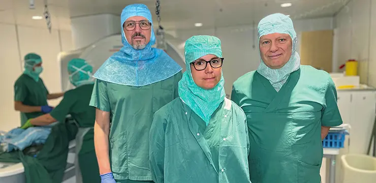
- The LITT treatments were performed in a modified clinical MRI scanner at Skåne University Hospital. By constructing a sterile “tent” around the patient, conditions like those of a traditional operating theatre were created.
- Most side effects observed were mild and transient. They were not judged to be caused by the equipment used in the study. Of the 24 reported adverse events, eight involved neurological or cognitive symptoms — all of which were mild. Two of these remained after three months, while the others resolved spontaneously.