Bowel ultrasound an effective tool for assessing patients with chronic inflammatory bowel disease

Getting a diagnosis can be a lengthy process for someone with IBD, such as ulcerative colitis or Crohn’s disease. Delays can occur for several reasons. One is that symptoms – abdominal pain, diarrhoea, bloody stools, and fatigue – are not specific to IBD and can be seen in other gastrointestinal conditions. Another is that standard diagnostic tools like MRI or CT often involve long waiting times.
“Colonoscopy, MRI, and CT are all excellent diagnostic methods. However, colonoscopy can be uncomfortable, which may discourage some patients from seeking care,” says Tillmann Raith, consultant in gastroenterology at Skåne University Hospital.
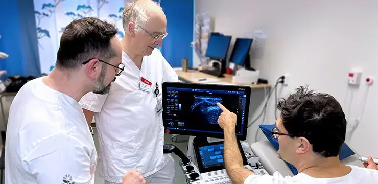
Together with colleagues Peter Gedeon, senior consultant, and Mladen Makitan, consultant, he is advocating for the wider use of an alternative method: bowel ultrasound. This technique uses ultrasound to detect signs of active inflammation or other IBD-related abnormalities in the intestines.
“We can examine patients directly in the clinic, using relatively simple equipment that doesn’t require extensive resources or much time. It’s a risk-free method that patients often appreciate as it’s quick and painless,” says Tillmann Raith.
Monitoring patients with IBD
At Skåne University Hospital, where the gastroenterology outpatient clinic in Malmö cares for around 5,000 patients with IBD, bowel ultrasound is routinely used to monitor disease activity.
“It’s an examination that can be repeated as often as needed. We’re available at short notice in the clinic and can assess patients quickly. The image quality with ultrasound has improved enormously over the past 15 years, which significantly enhances the reliability of our findings,” says Peter Gedeon.
He emphasises that the method is not intended to replace traditional diagnostic tools.
“Very superficial changes cannot be detected by ultrasound – for those cases, patients need to be assessed with capsule endoscopy or colonoscopy. Changes in the pelvic region are more clearly seen using MRI. But bowel ultrasound is an effective complement that can help expedite treatment,” says Peter Gedeon.